
《Published: 17 October 2025》
Haijiao Liu, Estela Noguera-Ortega, Xuanqi Dong, Won Dong Lee, Jeehan Chang, Sezin Aday Aydin, Yumei Li, Yonghee Shin, Xinyi Shi, Maria Liousia, Marina C. Martinez, Joshua J. Brotman, Soyeon Kim, Zeyu Chen, Anni Wang, Zirui Ou, Jungwook Paek, Ju Young Park, Aidi Liu, Haonan Hu, Zebin Xiao, Dora Maria Racca, Se-jeong Kim, G. Scott Worthen, …Dan Dongeun Huh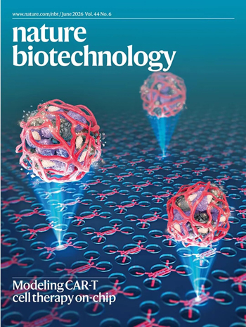
이번 호 Nature Biotechnology의 표지는 펜실베이니아 대학교 Dan Dongeun Huh 연구팀이 폐 면역학, 임상 종양학, 대사학 등 다학제 팀과 협력하여 수행한 연구 "A tumor‑on‑a‑chip for in vitro study of CAR‑T cell immunotherapy in solid tumors"를 주제로 합니다. 표지 이미지는 Haijiao Liu가 제작하고 Erin Dewalt가 디자인한 것으로, 혈관화된 종양-온-어-칩을 생생하게 묘사하여 고형 종양 미세환경의 복잡성을 실시간 고해상도로 분석할 수 있는 미세공학 시스템이라는 연구의 핵심 혁신을 상징합니다.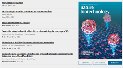
연구 배경
키메라 항원 수용체(CAR)-T 세포 치료는 혈액암에서 혁명적 성공을 거두었으나, 고형 종양 치료에는 여전히 막대한 난제가 존재합니다. 핵심 장애물은 종양 특이적 항원의 부족, 종양 미세환경(TME)이 가하는 물리적 장벽, 그리고 CAR-T 세포의 이동, 침윤, 기능을 저해하는 강력한 면역억제 신호 등입니다.
전통적 동물 모델은 인간 종양과 면역계 간의 복잡한 상호작용을 완전히 재현하지 못해, 전임상 결과와 임상 전환 사이에 심각한 간극이 존재합니다. 더욱이 암-면역 상호작용에 대한 제한된 이해는 고형암 대상 CAR-T 세포 치료 발전의 근본적 장벽으로 남아 있습니다.
연구 의의
본 연구는 인간 종양 절편의 혈관화와 면역 세포의 제어된 관류를 가능하게 하는 획기적 미세공학 시스템을 개발하여, 연구자들이 시험관 내에서 종양 미세환경 내 CAR-T 세포 활성을 모델링할 수 있게 했습니다. 주요 혁신은 다음과 같습니다.
1. 인간 고형 종양-면역 상호작용의 완전한 가시화를 가능하게 하는 "혈관화 종양-온-어-칩"
연구팀은 환자 유래 종양 절편 또는 PDX 조직을 관류 가능한 인간 혈관 네트워크가 포함된 미세공학 칩에 이식하였습니다. 14일 내에 이 시스템은 장기간 생존 가능하고 광학적으로 추적 가능한 혈관화 종양 모델을 형성했습니다. 처음으로 CAR-T 세포의 혈관 외 유출, 이동, 항원 인식, 사멸, 지속성의 전 과정이 시험관 내에서 재현되었으며, 고시공간 분해능 이미징 및 단일세포 시퀀싱을 통해 인간 고형 종양 면역 반응에 대한 실시간 정량 분석이 가능해졌습니다.
2. 두 가지 상승적 강화 전략—CCR2 "장갑화" 및 DPP4 억제—의 발견 및 분자 메커니즘 규명
칩-마우스 이중 모델 시스템을 통해 다음을 입증했습니다.
3. 표적 발굴 및 대사 바이오마커 식별을 위한 리간드-수용체 빅데이터 플랫폼 구축
scRNA-seq와 CellPhoneDB를 통합하여 150개 이상의 유의미한 상호작용을 확인했으며, 여기에는 CD38‑PECAM1, LTB‑LTBR, CXCL10/11‑CXCR3의 세 가지 주요 내피-CAR-T 경로가 포함됩니다. 항체 차단 실험은 이들 경로 중 어느 하나를 억제하더라도 CAR-T 주화성 및 항종양 효능이 유의미하게 손상됨을 확인했습니다. 한편, 칩 유출액의 비표적 대사체학은 7~9일 앞서 치료 반응을 예측할 수 있는 6가지 동적 바이오마커(예: 3-니트로티로신, 2-하이드록시부티레이트)를 식별하여, 향후 임상 모니터링을 위한 간단한 액체 생검 지표를 제공했습니다.
연구 전망
이 미세생리학 시스템은 암 및 기타 질환에 대한 세포 치료제 개발을 촉진할 유망한 시험관 내 플랫폼을 제공합니다. 향후 연구 방향은 다음과 같습니다.
표지 디자인 과정
표지 디자인은 "CAR-T 세포 치료의 칩 상 모델링"이라는 핵심 주제를 중심으로, "혈관화된 종양 구체 + 미세유체 칩 어레이"를 이중 시각적 앵커로 삼았습니다. 이미지 중앙의 세 개의 매달린 구형 구조는 "인간 종양 절편"을 직관적으로 나타내며, 표면을 감싸는 붉은 망상 네트워크는 "혈관 네트워크"를, 하단의 규칙적 배열 마이크로웰 어레이는 "미세유체 칩"의 물리적 지지체를 암시합니다. 이러한 조합은 논문 제목의 "종양-온-어-칩" 개념을 직접적으로 반영하여, 추상적 생체공학 시스템을 구체적 시각 형태로 구현했습니다.
표지는 연구에 사용된 인간 종양 절편을 대표하는 세 개의 매달린 구체로 구성됩니다. 각 구체 내부는 보라색과 검정색 입자 질감으로 실제 종양 세포 덩어리와 복잡한 세포외 기질을 모사하였고, 표면을 덮는 밝은 붉은 망상선은 종양 혈관 신생 과정을 생생히 재현했습니다. 이는 연구의 핵심 혁신인 종양의 혈액 공급 체계를 시험관 내에서 재건하여 면역 세포가 혈관을 통해 종양 내부로 진입할 수 있게 한 점을 보여줍니다. 구체 주변에 흩뿌려진 작은 빛 점들은 세포 활동과 미세 규모의 동적 반응을 암시합니다.
배경은 미세유체 칩의 대표적 구조인 진한 파란색 마이크로웰 어레이로, 고처리량 스크리닝과 정밀 제어된 실험 환경을 나타냅니다. 마이크로웰은 가는 빨간 선으로 연결되어 있으며, 이는 칩 내 유체 채널과 면역 신호 또는 CAR-T 세포의 전체 시스템 내 흐름 및 상호작용을 상징합니다. 하단 마이크로웰에서 위로 뻗은 원뿔형 파란색 광선은 공간적 깊이감을 줄 뿐만 아니라 칩 기술이 종양 모델에 제공하는 "지원"과 "역량 강화"를 상징하며, 생물학 연구에서 공학의 주도적 역할을 부각합니다. 이 디자인은 생물학의 복잡성과 공학의 정밀성을 동시에 보여주며, "시험관 내에서 생체 내 환경을 모사"한다는 연구의 본질을 완벽하게 포착했습니다. 이 디자인은 저널 편집자들의 인정을 받아 표지로 선정되었습니다!
Anhui Province Academy of Science and Technology, No. 128,Wanshui Road, Hi-Tech Zone, Hefei City, Anhui Province, China
 +86 17354022649
 service@sondii.com


영업 시간
11월 21일 월요일 - 11월 23일 수요일: 오전 9시 - 오후 8시
11/24 목요일: 휴무 - 즐거운 추수감사절 보내세요!
11월 25일 금요일: 오전 8시 - 오후 10시
11월 26일 토요일 - 11월 27일 일요일: 오전 10시 - 오후 9시
(모든 시간은 동부 표준시 기준)
 IPv6 네트워크 지원