
Research visualization and journal cover design can do more than make a scientific paper visually appealing.
A well-designed scientific cover can translate complex technologies into an intuitive visual story, helping readers quickly understand the connection between the research problem, technological innovation, and potential clinical impact.
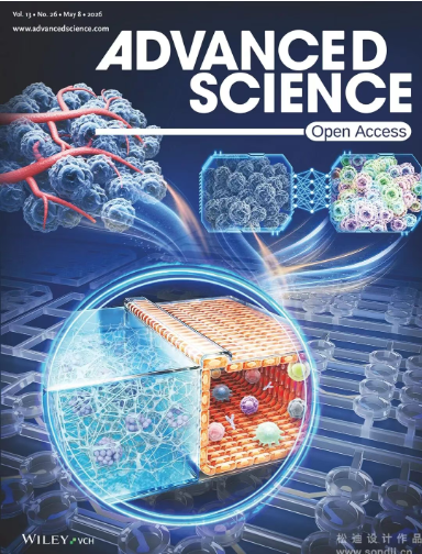
In Advanced Science Volume 13, Issue 26, the cover features the research article “Deep Learning-Powered Scalable Cancer Organ Chip for Cancer Precision Medicine,” authored by a multidisciplinary team from Harvard Medical School, the Shenzhen Institute of Advanced Technology, the Henan Academy of Medical Sciences, and other institutions.
The study presents an innovative combination of cancer organ-on-chip technology, patient-derived cells, microfluidics, and deep learning, offering a scalable approach to functional precision oncology.
For this project, the cover artwork was designed to visually communicate how an AI-powered organ-chip platform can bridge patient biology, high-throughput drug testing, and precision medicine.
Cancer treatment is becoming increasingly personalized. However, selecting the right therapy for an individual patient remains challenging.
Traditional patient-derived models, including patient-derived xenografts (PDX) and patient-derived organoids (PDO), can provide valuable biological information, but their clinical application is often limited by several practical challenges:
Complex and difficult-to-standardize experimental procedures
High cost
Long experimental cycles
Limited throughput
Incomplete recreation of the tumor microenvironment
Challenges in quantitative and longitudinal drug-response assessment
These limitations create a critical gap between laboratory models and real-world clinical decision-making.
An ideal platform for functional precision oncology should therefore combine biological fidelity, scalability, automation, quantitative analysis, and clinically relevant drug-response prediction.
The research team addressed this challenge by developing a scalable cancer organ-on-chip platform integrated with deep learning-based image analysis.
The study developed a fully thermoplastic injection-molded cancer organ-chip platform designed for scalable and automated experimentation.
One of its key innovations is a patented channel architecture combined with surface treatment technology that enables membrane-free and post-free capillary-pinned hydrogel confinement.
This configuration provides several advantages.
The platform can support:
Tissue-specific extracellular matrix environments
Tumor and immune cell co-culture
Automated experimental workflows
High-resolution imaging
High-throughput drug screening
Quantitative phenotypic analysis
By integrating these capabilities into a standardized chip platform, the researchers created a system that is better suited to repeated and scalable drug-response testing.
Importantly, the platform was validated using both cancer cell lines and patient-derived primary cells, demonstrating its potential for personalized drug sensitivity testing.
One of the most interesting aspects of this research is the integration of deep learning into the organ-chip workflow.
Conventional fluorescence imaging can provide valuable information about cellular phenotypes, but repeated fluorescent labeling can increase experimental complexity and may interfere with longitudinal observations.
To address this challenge, the researchers incorporated a deep learning-based image transformation model.
The system can use bright-field images to predict fluorescence-like information, allowing researchers to extract high-value phenotypic information without relying entirely on conventional fluorescent staining.
This creates an AI-assisted imaging workflow:
Bright-field imaging → Deep learning image transformation → Phenotypic information → Drug-response analysis
Such an approach has the potential to make longitudinal drug-response monitoring more efficient while reducing the dependence on fluorescent labeling.
The combination of microfluidics + patient-derived cells + high-resolution imaging + artificial intelligence therefore creates a powerful closed-loop system for functional precision oncology.
The significance of this technology goes beyond the development of another organ-chip platform.
The researchers demonstrated that the system could be used to evaluate drug sensitivity in patient-derived cells, with quantitative phenotypic results showing strong agreement with clinical therapeutic outcomes.
This suggests a potential future workflow in which patient-derived tumor cells could be introduced into a standardized organ-chip system, exposed to different therapeutic strategies, and analyzed through automated imaging and AI-assisted phenotypic interpretation.
In simplified form, the concept can be understood as:
Patient-derived cells → Cancer organ chip → Drug treatment → Imaging → AI analysis → Drug-response prediction → Personalized treatment decision
This closed-loop approach could help move functional precision oncology closer to practical clinical implementation.
For a research project combining cancer biology, microfluidics, organ-on-chip technology, artificial intelligence, and precision medicine, the challenge of cover design was not simply to illustrate individual components.
The key challenge was to visually communicate how these technologies work together as an integrated system.
Our design concept was therefore built around the visual metaphor:
The composition uses a three-level nested structure:
Macroscopic tumor → Mesoscopic organ chip → Microscopic AI
This visual hierarchy allows the viewer to move from the biological problem to the technological platform and finally to the intelligent analysis system.
The upper-left area of the composition presents a blue tumor mass surrounded by a red vascular network.
This element represents the complex biological environment in which cancer develops and responds to treatment.
The contrast between the blue tumor tissue and red vascular structures creates an immediate visual association with:
Cancer → Tumor microenvironment → Biological complexity
Rather than depicting cancer as an isolated cellular structure, the illustration emphasizes the surrounding biological environment, reflecting the research's focus on creating more physiologically relevant cancer models.
At the center of the composition, two floating hexagonal chip modules represent the organ-on-chip platform.
The left module presents an untreated cellular state, while the right module shows a transformed and differentiated cellular phenotype following computational analysis.
The two modules create a visual “before-and-after” comparison.
This helps communicate the central idea of the research:
Biological information enters the system → AI processes the information → Quantifiable phenotypic information emerges
The hexagonal geometry also echoes the structured architecture of microfluidic devices while giving the central composition a modern technological identity.
The transition from the biological state to the computationally analyzed state is represented through a neural-network-inspired visual transformation.
Rather than directly drawing an abstract AI brain or conventional circuit board, the design uses the transformation of cellular information itself to represent artificial intelligence.
This approach keeps the visual language closely connected to the actual research.
The AI is therefore not presented as an independent technology.
Instead, it is shown as an analytical layer operating on biological data.
This distinction is important because the research is fundamentally about integrating AI with experimental biology rather than simply applying AI as a standalone computational tool.
The circular enlarged section at the bottom provides a detailed view of the organ-chip architecture.
The illustration shows:
An orange epithelial layer
A blue extracellular matrix region
Tumor cells
Immune cells
Microfluidic channels
Culture wells
Cellular interactions within the chip
This magnified section serves two purposes.
First, it allows readers to understand the physical structure of the organ-chip platform.
Second, it introduces the biological complexity hidden inside the seemingly simple chip.
The contrast between the orange cellular layer and blue matrix region creates clear spatial separation, making the microenvironment easier to understand even at a glance.
Another important aspect of the study is its emphasis on scalability and automation.
To communicate this, the lower section incorporates repeated culture wells and surrounding microfluidic structures.
The repeated geometry suggests:
Standardization → Parallelization → High throughput → Automated analysis
This visual language is particularly important because scalability is one of the major differences between a promising laboratory prototype and a technology with potential for broader clinical or pharmaceutical applications.
Instead of showing automation through generic robotic imagery, the cover embeds the concept directly into the architecture of the organ-chip system.
The overall palette combines deep blue with orange-red accents.
Blue is used to communicate:
Technology
Data
Artificial intelligence
Precision
Computational analysis
Orange and red introduce:
Cellular activity
Blood vessels
Biological processes
Energy
Life
The interaction between these colors reinforces the central message of the study:
A light beam radiating outward from the center of the chip further symbolizes the flow of information and the potential of data-driven technologies to enable precision medicine.
One of the biggest challenges in scientific illustration is deciding what to show and what to leave out.
This research involves multiple layers of information:
Cancer biology + patient-derived cells + organ-on-chip + microfluidics + drug screening + imaging + deep learning + precision medicine
If every component were displayed with equal visual weight, the cover could quickly become overcrowded and difficult to understand.
Instead, the design establishes a clear hierarchy:
The biological problem.
The experimental platform.
The intelligent analysis layer.
The ultimate application.
This transforms a complex multidisciplinary research story into a visual narrative that can be understood within seconds.
The potential of this technology extends beyond the current study.
Future research could explore the application of the platform to additional cancer types, including:
Lung cancer
Breast cancer
Colorectal cancer
The platform could also be expanded to investigate combination therapies and more complex treatment strategies.
At the same time, integrating organ-chip experiments with technologies such as:
Single-cell sequencing
Spatial transcriptomics
Multi-omics analysis
AI-based image analysis
could provide researchers with increasingly comprehensive views of tumor biology.
Further standardization and modularization of chip manufacturing may also help accelerate the transition from laboratory research to practical applications in hospitals, pharmaceutical development, and third-party testing laboratories.
In the long term, the convergence of AI + microfluidics + patient-derived cells could establish a new technological infrastructure for precision oncology.
The Advanced Science cover illustrates an important principle of scientific communication:
A strong scientific illustration does not simply reproduce the experiment. It reveals the logic behind the experiment.
For this project, the visual story connects four key concepts:
Tumor biology → Organ-on-chip technology → AI-powered analysis → Precision medicine
By organizing these elements into a clear visual hierarchy, the cover communicates both the technical innovation and the clinical vision of the research.
We are proud that this cover design was recognized by the journal editors and successfully published in Advanced Science, Volume 13, Issue 26.
For researchers developing innovative technologies in biomedicine, cancer research, organ-on-chip systems, AI, microfluidics, or precision medicine, scientific visualization can help transform complex research into a compelling and accessible visual story.
Want to turn your research into a publication-ready scientific illustration or journal cover?
Our scientific illustration team can help translate complex research concepts into clear, engaging, and publication-ready visuals—from scientific figures and graphical abstracts to journal covers and research animations.
Anhui Province Academy of Science and Technology, No. 128,Wanshui Road, Hi-Tech Zone, Hefei City, Anhui Province, China
 +86 17354022649
 service@sondii.com


영업 시간
11월 21일 월요일 - 11월 23일 수요일: 오전 9시 - 오후 8시
11/24 목요일: 휴무 - 즐거운 추수감사절 보내세요!
11월 25일 금요일: 오전 8시 - 오후 10시
11월 26일 토요일 - 11월 27일 일요일: 오전 10시 - 오후 9시
(모든 시간은 동부 표준시 기준)
 IPv6 네트워크 지원